Cardiac catheterisation and angiography
Angiography is a procedure that uses an injection of a liquid dye to watch blood flow through the arteries that supply your heart muscle (coronary arteries). It can also give information about the pressures and function of the ventricles.
The procedure takes place in an x-ray room and takes from 20 minutes to an hour, depending on what is found.
A team of healthcare professionals are involved in the procedure, including a doctor, nurse, technician and radiographer.
A catheter is passed into a vein or artery, either in your groin or arm. You will be given a local anaesthetic so you will not feel this. X-ray screening is used to help direct the catheter through your blood vessels and into the correct position in your heart. You won’t feel the catheter moving and can choose to watch it on the video screen.
Once in position, the blood pressure at the tip of the catheter will be checked. Then a dye will be put down the catheter and a series of x-ray pictures will be taken.
When the procedure is over the catheter is removed and a nurse will apply a dressing.
After the test you will have to rest for several hours and you may find you feel fatigued for a while. The place where the catheter was inserted may be sore and you may have a small bleed or small lump around the area, but this will disappear after a few days.
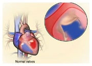
Angiography gives vital information about the pressures inside your heart, how well your heart is working and the blood flow in your coronary arteries. This procedure also enables the cardiologist to assess if you have poorly functioning valves.
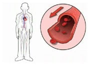
It can also locate narrowing of the arteries that supply blood to your heart muscle, and determine how serious they are.
The results of an angiography will assist in decisions about possible interventions or surgery.


