Chest x-ray
A Chest x-ray is a type of photograph of the heart, lungs, blood vessels and the bones of the spine and chest. It doesn’t show specific details of the heart and so only general changes in shape and size can be seen.
You will have to visit the radiology department in your hospital. There, you will be asked to stand with your chest pressed up against a photographic plate and to take a deep breath and stand still. The test is painless and harmless and the amount of radiation is small – but the photographic plate may be cold which could be uncomfortable. A radiographer will then press a button that sends a beam of X-rays to the photographic plate.
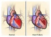
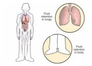
After a short time, your X-ray will be ready. The doctor will be able to see if your heart is enlarged or if there are any signs of congestion or infection. In addition, the chest X-ray may reveal lung diseases, which may cause symptoms like those experienced by patients with heart failure.

