Multi-slice computer tomography (MSCT)
Cardiac imaging by multi-slice computed tomography (MSCT) is a recently developed technique for assessing the function of the heart and coronary arteries non-invasively. Cardiac MSCT uses X-ray beams and a liquid dye to form a 3-D image of the heart and vessels. The scanning machine used is very sophisticated and scans the heart very quickly. This provides sharp, detailed images that can’t be achieved with other tests. In the same way as if you were to have an angiogram, you will be injected with a liquid dye to look for any narrowing of your coronary arteries.
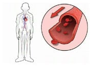
In contrast to coronary angiography, where the liquid dye is injected by an invasive catheter into your coronary arteries directly, the liquid dye during this examination is injected into a superficial vein by a small needle placed either on the back of one of your hands or in your elbow groove. Your body then transports the dye when circulating your blood and the examination will start when it reaches your coronary arteries.
A CT scanner uses X-rays to scan the dye moving through your heart and its blood vessels to create sharp, detailed images. The examination only takes a few seconds and is usually done during a short breath hold. The amount of radiation you are exposed to is very low.
Return to Common tests for heart failure


